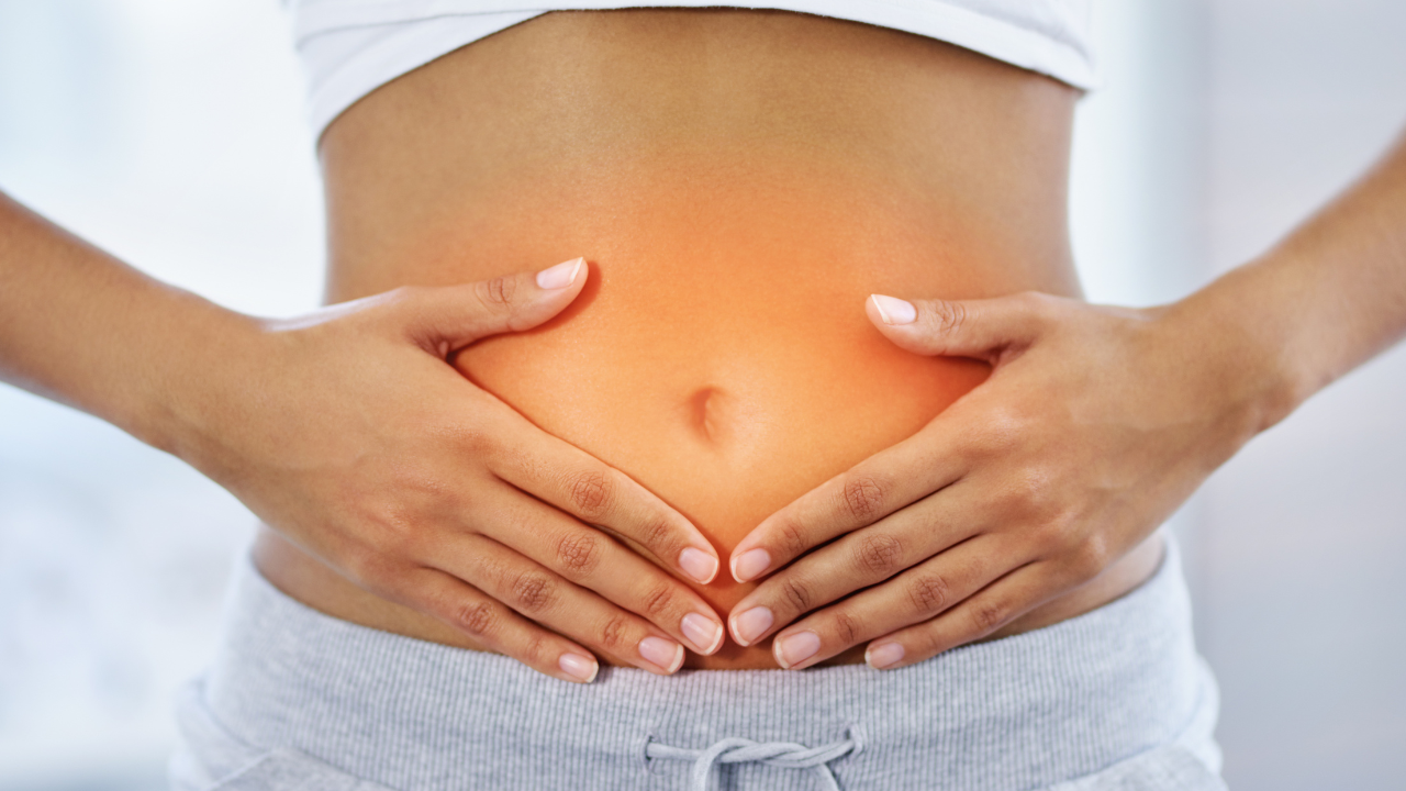
The Fascinating Connection Between Liver Disease, SIBO and Your Gut Health
Non-alcoholic fatty liver disease (NAFLD) is the most common liver disease, affecting one in four adults globally, and approximately 7.8 million Canadians [1]!
With the growing number of cases around the world, there has been greater interest and research on this condition, with one of the growing focuses being the gut-liver relationship.
Join us in today’s post where we investigate non-alcoholic fatty liver disease, the relationship between gut health and liver disease, and dietary changes to support a healthy liver.

Functions of the Liver
Did you know - the liver is one of the largest organs in the body?
It is involved in the metabolism of carbohydrates, protein, and fat; micronutrient conversion and/or storage of vitamin A, vitamin D, water soluble vitamins, iron, and zinc; and detoxification of drugs, alcohol, and toxic substances like ammonia and urea.
The liver has enormous regenerative capacity that allows the liver to continue to perform its functions and responsibilities despite disease. It is only under larger damage that the regenerative capacity is exceeded, and signs and symptoms of disease begin to appear.

What is NAFLD?
There are two main forms of fatty liver disease: alcohol-induced fatty liver disease (AIFLD) and non-alcoholic fatty liver disease (NAFLD). AIFLD is caused by heavy drinking, while NAFLD occurs in individuals who do not drink excessively.
It is estimated that the prevalence of NAFLD in Canada will increase by 20% between 2019 and 2030 [1]. NAFLD specifically refers to the conditions that are a result of an accumulation of fat in the liver. NAFLD develops in stages beginning from less severe to very severe, and it can take years for individuals to reach the last two stages.
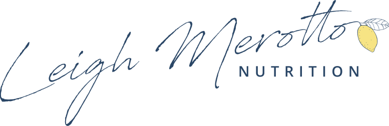
There are not many symptoms during the first two stages of NAFLD, however, individuals in the later stages of NAFLD typically experience a dull ache in the stop stomach, extreme fatigue, and unexplained weight loss.
The four stages are:
Steatosis aka simple fatty liver - is a small build-up of fat in the liver. This build up of fat occurs because excess fat is presented to the liver, or there is a reduction in the liver’s ability to export fat. At this stage, there are little to no symptoms, but individuals may experience fatigue, anorexia, nausea, and vomiting. The damage is reversible at this stage of liver disease.
Non-alcoholic steatohepatitis (NASH) - is fatty liver (steatosis) with associated inflammation. Symptoms are similar to those for steatosis, but may also include fever and weight loss. At this stage, damage can still be reversible, but if damage continues it may lead to liver failure, hepatic encephalopathy, or cirrhosis.
Fibrosis - persistent inflammation resulting in the development of scar tissue around the liver and nearby blood vessels.
Cirrhosis - end stage of liver disease where excess scar tissue causes the liver to shrink due to death of liver cells. The scar tissue eventually replaces the healthy liver tissue, and this damage is permanent and can slow the functions of the liver as the scar tissue blocks the flow of blood through the liver. Common symptoms of this stage include jaundice (yellowing of the skin and whites of the eyes), itchy skin, and swelling of the legs, ankles, feet or stomach due to excess water retention. Long-term cirrhosis can cause liver failure or liver cancer.

Diagnosis
Most physicians use a combination of your medical history, a physical/clinical exam, and diagnostic tests in order to make an NAFLD diagnosis.
Although it is unknown what the exact cause(s) of NAFLD is, there are known risk factors that can increase your chances of developing NAFLD. These risk factors are often included in your medical history and will be used during the diagnosis process.
Risk factors could be:
Being obese or overweight
Being diagnosed with type 2 diabetes
Having high blood pressure
Being older than the age of 50
Smoking
Having high cholesterol

Clinical manifestations (aka connected symptoms and conditions) of NAFLD include:
Portal hypertension: high blood pressure in the vein (known as the portal vein) that brings blood from the gastrointestinal tract and spleen to the liver. During the last few stages of NAFLD, the scar tissue and dead liver cells create blockages that increase the resistance of blood flow through the portal vein. This raised resistance increases the blood pressure within the portal vein and results in the blood travelling through smaller, alternate channels. As a result of portal hypertension, there are less nutrients being delivered to the liver, and affects the various functions of the liver.
Ascites: this condition is associated with portal hypertension, where the raised blood pressure causes blood pressure in the smaller, alternate channels around the internal organs of the body to increase as well. As the blood pressure rises, fluid leaks into the peritoneal cavity (cavity within the abdomen that is sandwiched between a membrane that covers your abdomen and pelvis, and the other membrane that covers your internal organs). As more and more fluid drains into the peritoneal cavity, you develop ascites, where the excess fluid builds up, creating the appearance of a ‘beer belly’.
Jaundice: is more easily detected as it causes the yellowing of the skin and whites of eyes. It is a result of an accumulation of total bilirubin (waste products of the normal breakdown of red blood cells) the extracellular fluid. Individual’s may also experience itchy skin (aka pruritus).
Esophageal varices: is the dilated and abnormal veins that occur in the lower part of the esophagus and often the upper part of the stomach. This is also caused by portal hypertension, as the increased blood flow in the smaller blood vessels leading to the esophagus, results in the enlargement of these vessels. This is often seen in more advanced NAFLD, and is very painful and may result in excessive bleeding.
Edema: is the build-up of fluid in the extravascular space, resulting in retention of water in the feet and legs.
Hepatic encephalopathy: since the liver is involved in the detoxification of the blood, poor liver function can result in the accumulation of toxic substances within the blood. This can cause delirium and poor ability in controlling muscular movement.
Osteoporosis/osteopenia: as the liver is involved in the metabolism of calcium, phosphorus, and vitamin D, poor liver function can cause osteoporosis or osteopenia. Osteopenia is the weakening of the bone, increasing its susceptibility to breakage. Osteoporosis is a more severe condition of osteopenia.

Common diagnostic tests include an endoscopy, ultrasound, or biopsy.
An endoscopy may be used to detect esophageal varices. It is a long, flexible tube with a camera at the end, that is inserted into the mouth through your esophagus to search for dilated and abnormal veins.
An ultrasound utilizes a transducer (wand) to create images of organ structures. This is used to detect enlargement of the liver.
Liver biopsy may be used which is the most invasive but the most accurate diagnostic test that also indicates the severity of liver damage. This process involves taking small portions of your liver and examining it under a microscope for signs of damage or disease.

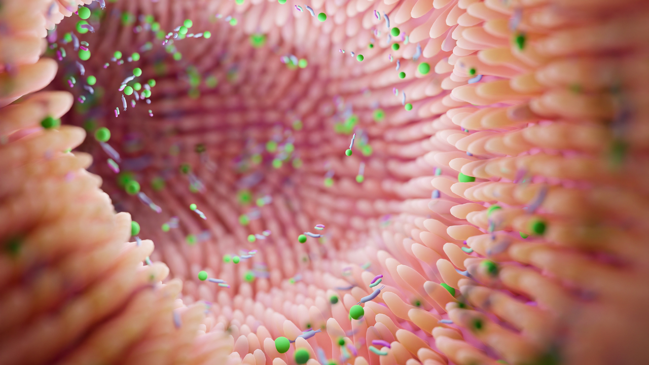
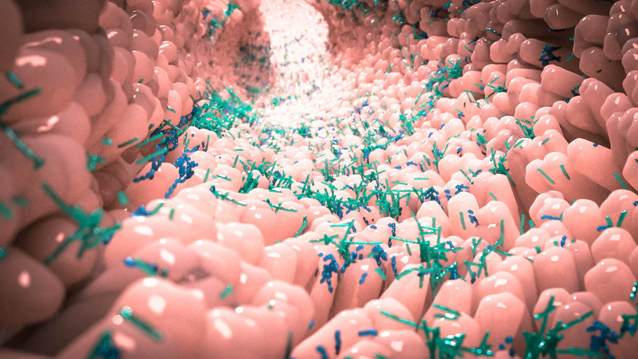
What is Gut-Health?
The gut is all the organs and structures from the mouth to the anal canal. The gut is involved in digestion, absorption, and excretion of food.
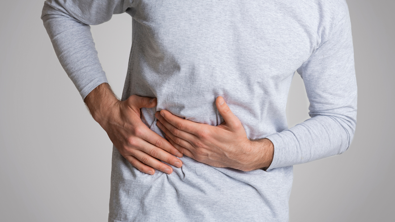
Our gut also houses our microbiomes (including bacteria, archaea, fungi, and viruses) that play a role in our mental health, mood, metabolism, digestion, absorption, and metabolism.
To have a healthy gut, we need a diverse microbiome. A greater variety of microbiomes supports the function of the gut and also promotes well-being as it reduces inflammation and lowers the risk of heart disease and obesity [2]. Amazing, huh?
You may want to read: Gut Microbiome: What Is It and How Can We Better Support Our Gut Bugs?
Gut microbiomes are influenced by a variety of factors such as age, disease, dietary habits, stress, alcohol intake, certain medications, etc [2]. Emerging studies are indicating that dietary factors may exhibit the greatest impact on gut health [2].
Since we know that inflammation is a key driver of liver damage and can result in a variety of negative impacts, as listed above. Continue to read, to learn more about the connection between the gut and NAFLD.
P.S. to learn more about gut microbiomes and gut health, be sure to follow along with me on Instagram!
The Gut-Liver Axis
The gut-liver axis is the close bi-directional relationship between the gut’s microbiome and the liver. These two systems interact and influence each other through the portal vein, which is the main vein supplying blood to the liver from the gastrointestinal tract and spleen. About 70% of the blood flowing from the portal vein into the liver comes from the intestines [3]! Each system closely affects each other, and damage to one system can result in damage to the other.
The liver secretes bile acids and IgA antibodies into the intestinal tract. Bile acids are substances that are developed in the liver and absorbed into the small intestine to aid in the digestion of fat; absorption of fat and fat-soluble vitamins; and excretion of cholesterol from the body.
Bile acids also contain immunoglobulin A (IgA) antibodies that are responsible for immunity by protecting the mucous membranes. Therefore, a reduction in bile acid secretion from the liver is associated with inflammation and microbial overgrowth in the gut [4].
Conversely, the liver receives nutrients, toxins, antigens, and small amounts of microbial components through this blood. It then removes pathogens or other potentially dangerous microbial components from the GI tract. It is the first organ, other than the intestines, to be exposed to gut derived products. With liver disease, pathogens from the gut can further cause liver damage due to poor liver function. The liver’s inability to detoxify blood from the GI tract can lead to small intestinal bacterial overgrowth (SIBO; more on this later!) or intestinal dysbiosis [3].
Dysbiosis of the gut microbiomes causes a steady, low level of chronic inflammation and results in the development of NAFLD. Dysbiosis is considered as a change in the composition of the gut microbiomes, where there may be less' good’ microbiomes and more ‘bad’ microbiomes.
Furthermore, since the two systems work symbiotically, changes in the composition of the gut microbiomes and bacterial overgrowth, can lead to a greater amount of microbiomes reaching the liver and causing damage to the liver. Studies have shown that individuals with NAFLD tend to have lower gut microbiome diversity [5].
Want my top 5 starter tips to reduce bloating and IBS symptoms, and support your microbiome? Grab my Beat the Bloat Cheat Sheet.

Fatty Liver Disease and SIBO
SIBO is small intestinal bacterial overgrowth and this condition is associated with an increase in the number of bacteria within the small intestine. An increase in the growth of bacteria within the small intestine can cause poor digestion and absorption function within the GI tract. (TIP: SIBO is a complex condition and can’t possibly be explained in just one paragraph, if you are interested in learning more about SIBO, check out this article!)
Emerging research has discovered that individual’s diagnosed with NAFLD have a significantly higher probability of also being diagnosed with SIBO, than those without NAFLD [6]. A study found that almost 35% of patients with NAFLD also have SIBO [7].
Knowing the gut-liver axis, and the relationship between the gut microbiomes and the liver, it is understandable how these two conditions are so tied together. There is evidence that those with NAFLD have an abnormal gut microbiome in comparison to those without NAFLD [7]. It is hypothesized that these abnormal microbiomes produce toxic substances to the liver and alter the liver’s ability to function and metabolize, ultimately contributing to the development of NAFLD [8].

It’s all making sense now, eh? A pretty fascinating connection, and if you have overlapping liver disease and gut issues like IBS and SIBO, hopefully this article has provided more insight as to why this is.
Some of the toxic substances produced due to SIBO that can contribute to the development of NAFLD are gram-negative bacteria. The literature has shown that SIBO results in the accumulation of gram-negative bacteria in the intestines [9]. These Gram-negative bacteria are extremely antibiotic resistant bacteria that can cause infections like pneumonia [10].
One of the outer membrane components of the gram-negative bacteria that makes the gram-negative bacteria so dangerous, is the lipopolysaccharide (LPS). The LPS can cause inflammation and damage to the intestinal barrier. As the LPS travels to the liver through the portal vein, it can cause inflammation within the liver and ultimately contribute to the development of steatosis and fibrosis of the liver [9].

Nutrition for a Healthy Liver
In the treatment for NAFLD, lifestyle methods are primarily used, including diet and physical activity. One of the most successful types of diets are those that follow the Mediterranean dietary pattern. Studies have shown that the combined effect of the Mediterranean diet (MD) and adhering to the recommended physical activity levels are more effective in treatment than a single pharmacological option [11].
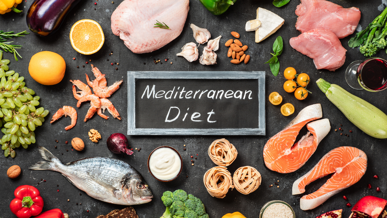
The MD diet is one that is high in plant-based foods, moderate consumption of protein-source foods, and low-to-moderate levels of red wine consumption, milk, meat, and dairy products.
A note about different cultural diets and the Mediterranean diet…
Although its name was coined due to its Mediterranean origins, its principles can work for a variety of cultural backgrounds and ethnic diets. It's not solely about eating Mediterranean foods, but a pattern of consuming more plant-based foods, while minimizing the consumption of meat and meat products, red wine, etc. Therefore, choose foods that fit into your lifestyle so that the dietary changes and choices that you make can have a long-term impact on your wellness.
Now back into how you can use these general principles to support your liver health.
Some of the results that were seen in individuals with NAFLD who followed the MD were decreased liver fat accumulation, triglycerides, cholesterol, and inflammatory biomarkers [12]. The reason why the MD may be so effective is because it encourages consumption of probiotics, prebiotics, fiber-rich foods, polyphenols, and antioxidants [12].
Since the mediterranean diet is a broad diet, here are some key nutrients to look out for:
Reduce consumption of saturated fatty acid (SFA). SFA’s are typically found in animal products such as red meats, butter, and dairy products, but can also be found in vegetable products like coconut oil and palm oil. Processed and sugary foods tend to be higher in SFA. These foods may be soft drinks, pre-packaged foods, red meats, sweet desserts, etc. Increased consumption of SFA is associated with worsening of NAFLD condition.
Consume a moderate amount of monounsaturated fatty acids (MUFA). MUFA are typically found in olive oil, avocado’s, palmitic acid, and nuts. MUFA contains polyphenols and phytochemicals which have antioxidant and anti-inflammatory functions. Therefore, consuming MUFAs may reduce steatosis in NAFLD.
Consume more omega-3 fatty acids. Omega-3s are typically found in seafood including salmon, tuna, cod, or shellfish; vegetable oils like flaxseed oil; eggs; and meat. Omega-3s improve the amount of liver enzymes and decrease triglyceride levels. Together, these PUFA produce beneficial effects in individuals diagnosed with NAFLD.
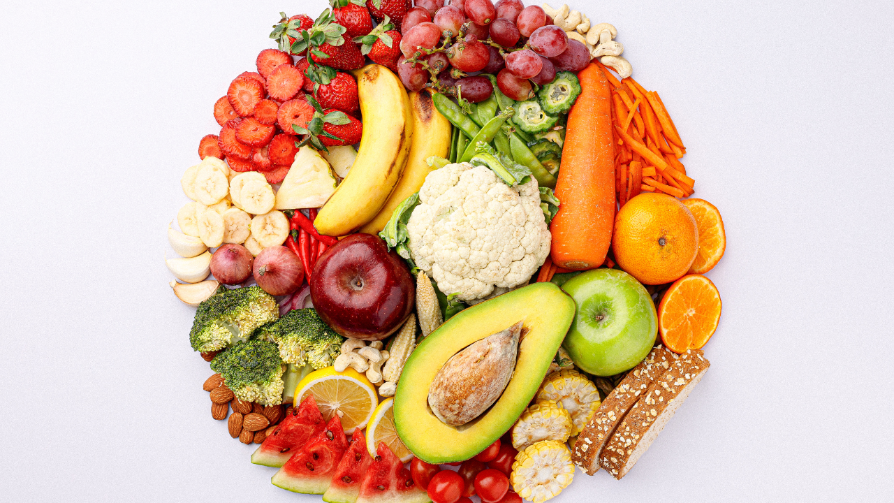
Consume more fiber-rich foods. Some fibers may be categorized as prebiotics. Prebiotics are parts of food that are indigestible and can act as food for the microbiomes in the gut. Some common prebiotic fibers are garlic, asparagus, leek, onion, and chicory root. Since they provide nutrients for our gut microbiomes, its positive impact in the gut may confer its benefits through the liver due to the gut-liver axis.
In summary, it is key to avoid pro-inflammatory foods like red meats, highly processed foods, and simple sugars. While it is important to incorporate more fiber-rich, omega-3s, and MUFA containing foods into your diet.
Want a head start on a Mediterranean diet? Grab my 7-Day Mediterranean Meal Plan here.

Final Thoughts
As we come to an end of this post, here are some key takeaways:
Liver disease is a progressive disease, and making lifestyle changes during the early stages, can prevent further progression and negative health outcomes
The gut and the liver is significantly intertwined, and poor gut health can impact the liver, and vice versa
There are many long-term diet changes that can support liver health. Limit pro-inflammatory foods, and instead choose high fiber, omega-3, and MUFA foods.
If you are feeling overwhelmed with all the information from this post, you’re not alone. NAFLD is a condition that is not even completely understood by researchers.
However, working with a Registered Dietitian for personalized guidance, help with overcoming mindset blocks and ongoing accountability can be a game changer. If you are interested in working together, apply for 1-1 coaching with me here.

Subscribe to our newsletter for new articles and the top gut-health & fitness tips.

BE SURE TO FOLLOW ME HERE
References:
[1] Sebastiani, G., Keyur, P., Vlad, R., Jordan, F., Neuschwander-Tetri, B., Pinzani, M., Petta, S., Berzigotti, A., Metrakos, P., Shoukry, N., & Brunt, E. (2022). Current considerations for clinical management and care of non-alcoholic fatty liver disease: Insights from the 1st International Workshop of the Canadian NASH Network (CanNASH). Canadian Liver Journal, 5(1), 61–90. https://doi.org/10.3138/canlivj-2021-0030
[2] Turnbaugh, P. J., Ridaura, V. K., Faith, J. J., Rey, F. E., Knight, R., & Gordon, J. I. (2009). The Effect of Diet on the Human Gut Microbiome: A Metagenomic Analysis in Humanized Gnotobiotic Mice. Science Translational Medicine, 1(6), 6ra14. https://doi.org/10.1126/scitranslmed.3000322
[3] Jasirwan, C., Lesmana, C., Hasan, I., Sulaiman, A., & Gani, R. (2019). The role of gut microbiota in non-alcoholic fatty liver disease: Pathways of mechanisms. Bioscience of Microbiota, Food and Health, 38(3), 81–88. https://doi.org/10.12938/bmfh.18-032
[4] Hersoug, L.-G., Møller, P., & Loft, S. (2016). Gut microbiota-derived lipopolysaccharide uptake and trafficking to adipose tissue: Implications for inflammation and obesity. Obesity Reviews: An Official Journal of the International Association for the Study of Obesity, 17(4), 297–312. https://doi.org/10.1111/obr.12370
[5] Rau, M., Rehman, A., Dittrich, M., Groen, A. K., Hermanns, H. M., Seyfried, F., Beyersdorf, N., Dandekar, T., Rosenstiel, P., & Geier, A. (2018). Fecal SCFAs and SCFA-producing bacteria in gut microbiome of human NAFLD as a putative link to systemic T-cell activation and advanced disease. United European Gastroenterology Journal, 6(10), 1496–1507. https://doi.org/10.1177/2050640618804444
[6] Mikolasevic, I., Delija, B., Mijic, A., Stevanovic, T., Skenderevic, N., Sosa, I., Krznaric-Zrnic, I., Abram, M., Krznaric, Z., Domislovic, V., Filipec Kanizaj, T., Radic-Kristo, D., Cubranic, A., Grubesic, A., Nakov, R., Skrobonja, I., Stimac, D., & Hauser, G. (2021). Small intestinal bacterial overgrowth and non-alcoholic fatty liver disease diagnosed by transient elastography and liver biopsy. International Journal of Clinical Practice, 75(4), e13947. https://doi.org/10.1111/ijcp.13947
[7] Gudan, A., Jamioł-Milc, D., Hawryłkowicz, V., Skonieczna-Żydecka, K., & Stachowska, E. (2022). The Prevalence of Small Intestinal Bacterial Overgrowth in Patients with Non-Alcoholic Liver Diseases: NAFLD, NASH, Fibrosis, Cirrhosis—A Systematic Review, Meta-Analysis and Meta-Regression. Nutrients, 14(24), Article 24. https://doi.org/10.3390/nu14245261
[8] Fei, N., Bruneau, A., Zhang, X., Wang, R., Wang, J., Rabot, S., Gérard, P., & Zhao, L. (2020). Endotoxin Producers Overgrowing in Human Gut Microbiota as the Causative Agents for Nonalcoholic Fatty Liver Disease. MBio, 11(1), e03263-19. https://doi.org/10.1128/mBio.03263-19
[9]Ghoshal, U. C., Baba, C. S., Ghoshal, U., Alexander, G., Misra, A., Saraswat, V. A., & Choudhuri, G. (2017). Low-grade small intestinal bacterial overgrowth is common in patients with non-alcoholic steatohepatitis on quantitative jejunal aspirate culture. Indian Journal of Gastroenterology, 36(5), 390–399. https://doi.org/10.1007/s12664-017-0797-6
[10] Gram-negative Bacterial Infection. (n.d.). Retrieved February 22, 2023, from https://www.winchesterhospital.org/health-library/article?id=936788
[11] Abenavoli, L., Milic, N., Di Renzo, L., Preveden, T., Medić-Stojanoska, M., & De Lorenzo, A. (2016). Metabolic aspects of adult patients with nonalcoholic fatty liver disease. World Journal of Gastroenterology, 22(31), 7006–7016. https://doi.org/10.3748/wjg.v22.i31.7006
[12] Suárez, M., Boqué, N., Del Bas, J. M., Mayneris-Perxachs, J., Arola, L., & Caimari, A. (2017). Mediterranean Diet and Multi-Ingredient-Based Interventions for the Management of Non-Alcoholic Fatty Liver Disease. Nutrients, 9(10), 1052. https://doi.org/10.3390/nu9101052